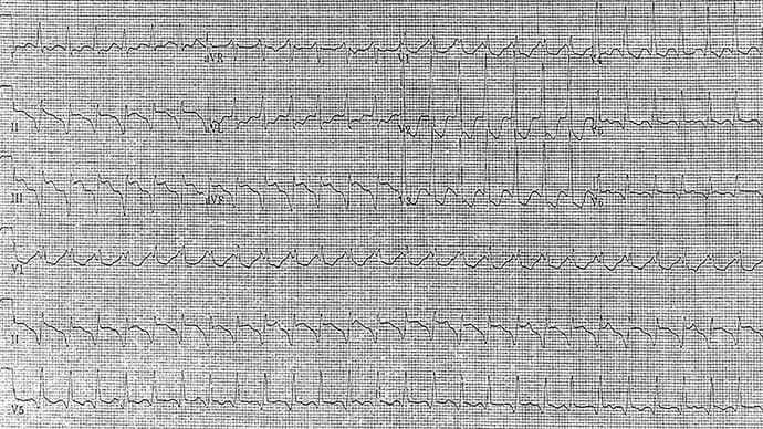
A 74-year-old man with a history of a myocardial infarction has sudden palpitations and presyncope for which he calls emergency medical services. He is brought to the emergency department, where his blood pressure is 80/50 mm Hg. His pulse is rapid and a 12-lead ECG is obtained.

Figure 1. Courtesy of Philip J. Podrid, MD.
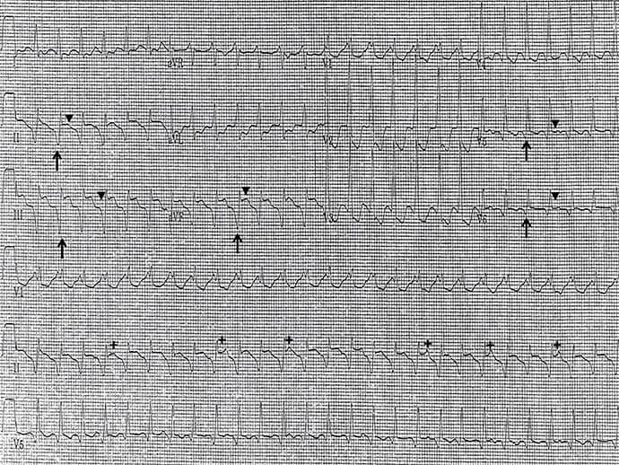
The correct diagnosis is ventricular tachycardia (Figure 2).

Figure 2. Courtesy of Philip J. Podrid, MD.
Discussion
The rhythm is regular at a rate of 160 beats/min. The QRS complex duration is increased (0.14 sec). The morphology resembles neither a left nor a right bundle branch block.
Q waves occur in leads II, aVF, and V5-V6 (↑). The ST segment is also elevated in these leads (▼). In addition, there is positive concordance (ie, tall R waves from V1-V6). There are subtle differences in the QRS complex morphology, especially in lead V1. Finally, P waves (+) occur (with a regular rate of 58 beats/min) that are dissociated from the QRS complexes.
A number of features establish this rhythm as ventricular tachycardia: atrioventricular dissociation, variability of QRS complex morphology, and positive concordance across the precordium. The presence of Q waves in the inferior and anterolateral leads suggests that the tachycardia originates in the inferolateral wall, probably a result of a prior inferior wall infarction and scar. The ST segment elevation likely reflects the development of a wall motion abnormality.
Philip Podrid, MD, is an electrophysiologist, a professor of medicine and pharmacology at Boston University School of Medicine, and a lecturer in medicine at Harvard Medical School. Although retired from clinical practice, he continues to teach clinical cardiology and especially ECGs to medical students, house staff, and cardiology fellows at many major teaching hospitals in Massachusetts. In his limited free time he enjoys photography, music, and reading.

